|
A 53 year-old man developed the acute onset of tetraplegia. On exam, he had paralysis of the tongue and flaccid weakness of all four extremities. Sensation to pinprick was absent below the neck, but vibration was relatively intact. |
![]()
![]()
![]()
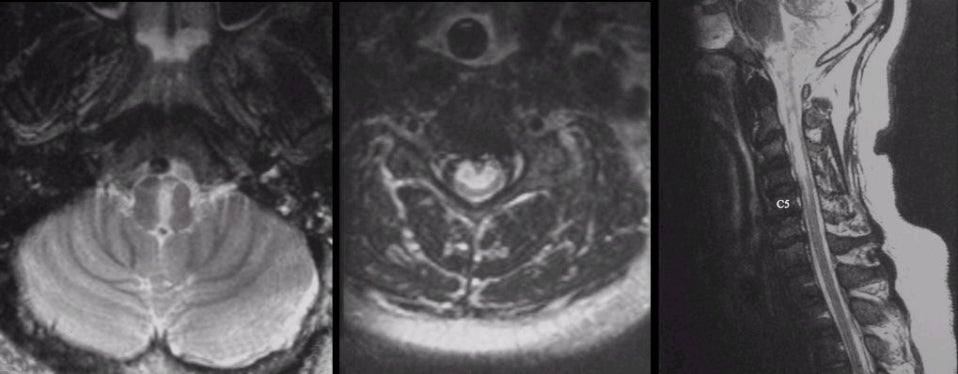
| Anterior Spinal Artery Stroke:
(Left) T2-weighted
axial MRI of the lower medulla; (Middle) T2-weighted axial MRI of the mid-cervical
spine; (Right) T2-weighted sagittal MRI of the upper cervical spine and lower
brainstem. On the left image, note the infarct in the medial medulla. On the middle image, note the infarct within the
anterior spinal cord, sparing the
posterior columns. On the right image, note the intramedullary
lesion extending from C5-C6 up through the medial medulla. This is a complete anterior
spinal artery (ASA) stroke.
The ASA arises from the intracranial vertebral artery where it supplies the medial medulla. It then descends on the anterior surface of the spinal cord supplying the anterior two-thirds of the spinal cord. Radicular arteries throughout the spine also anastomose with the ASA. Complete infarctions result in a complete spinal cord syndrome with the exception of the posterior columns, which are spared. Hence, the sparing of vibration. The posterior columns are supplied by the posterior spinal arteries. |
Revised
11/29/06
Copyrighted 2006. David C Preston