|
A 23 year-old woman developed the sudden onset of a severe headache. |
![]()
![]()
![]()
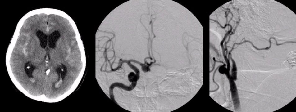
| Subarachnoid Hemorrhage - Anterior Communicating Artery
Aneurysm and Fibromuscular Dysplasia (FMD). (Left)
Axial CT
scan without contrast; (Middle)
Right internal carotid
artery angiogram, AP view; (Right) Common carotid artery angiogram. On the CT scan, note the subarachnoid blood in the sulci and posterior horns of the lateral ventricles.
On the internal carotid artery angiogram, the anterior communicating artery aneurysm
(the source of the bleeding) is clearly seen. Furthermore, note that on
the common carotid artery injection, the internal carotid artery is narrow and beaded. This is the picture of fibromuscular dysplasia. ICA = internal carotid artery, MCA = middle cerebral artery, ACA =
anterior communicating artery, CCA = common carotid artery, ECA = external carotid artery. FMD is a disease of the arteries which often affects the internal carotid arteries (presenting as TIA, stroke or intracranial aneurysms) or the renal arteries (presenting as renovascular hypertension). Other affected arteries include the vertebral, lumbar, mesenteric, celiac, hepatic, and iliac arteries. FMD occurs most commonly in young and middle-aged women. The etiology is unknown. Some studies have described an association of FMD with alpha-1 antitrypsin deficiency. Other proposed etiologies include hormonal effects on smooth muscle, mechanical stress on the arterial wall of affected arteries, and mural ischemia in dysplastic vessels. |
Revised
11/29/06.
Copyrighted 2006. David C Preston