|
An 85 year-old man presented with a headache, word finding problems and a right visual field loss. |
![]()
![]()
![]()
![]()
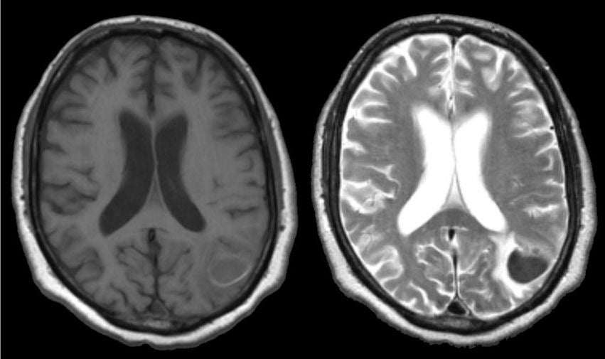
| Acute Intracerebral Hemorrhage: (Left) T1-weighted
axial MRI; (Right) T2-weighted axial MRI. Note that on the T1-weighted
scan, there
is an abnormality in the left parietal
lobe that is slightly hypointense. The same
area on the T2-weighted scan is dark with a surrounding bright signal.
This is the characteristic picture of an acute (approximately 3 days old) hemorrhage on MRI. In the acute stage, intracellular deoxyhemoglobin is dark on both T1- and T2-weighted scans. As the deoxyhemoglobin changes to intracellular methemoglobin, the signal becomes bright on T1-weighted images but remains dark on T2-weighted images. Thus, when this transition occurs, there is a time when the signal is isointense on T1-weighted scans. If one looks closely at the T1-weighted scan, one can see a faint rim of bright signal. This represents the early formation of intracellular methemoglobin. In this case, the hemorrhage was due to hypertension. The findings of blood on MRI are complex and depend on timing. To learn more, review the powerpoint slide show, Blood on MRI: Time-dependent Changes. |
Revised
11/29/06.
Copyrighted 2006. David C Preston.