|
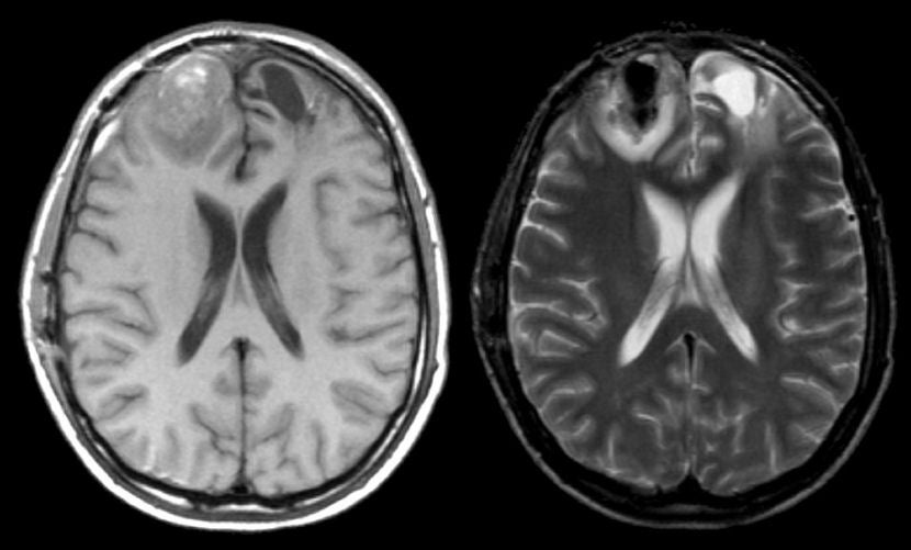
A 35 year-old man presented with a headache and confusion following a motor vehicle accident. |
![]()
![]()
![]()
| Acute Intracerebral Hemorrhage: (Left) T1-weighted
axial MRI; (Right) T2-weighted axial MRI. Note that on the T1-weighted
scan, there is an abnormality in the right frontal pole that is
mostly isointense. If one looks closely, one can see that there a hypointense
area inferiorly and a hyperintense area
superiorly. The same
area on the T2-weighted scan is dark with a surrounding bright signal. On
the left side, also note that there is edema in the frontal pole
(dark on T1-weighted and bright on T2-weighted images).
This is the characteristic picture of a hyperacute hemorrhage changing to an acute (approximately 3 days old) hemorrhage on MRI. The dark signal on T1- and T2-weighted scans represents deoxyhemoglobin whereas the isointense signal is oxyhemoglobin. The surrounding bright signal is vasogenic edema. The slight hyperintense signal on the T1-weighted scan is the early transition of some of the hemorrhage to intracellular methemoglobin. In this case, the hemorrhage was due to trauma. The findings of blood on MRI are complex and depend on timing. To learn more, review the powerpoint slide show, Blood on MRI: Time-dependent Changes. |
Revised
11/05/06.
Copyrighted 2006. David C Preston.