|
A 60 year-old man developed a headache followed by aphasia and right sided weakness. |
![]()
![]()
![]()
![]()
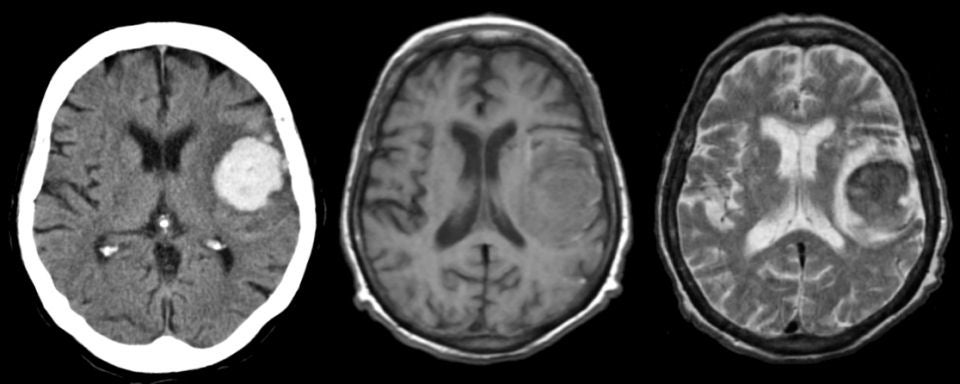
| Acute Intracerebral Hemorrhage: (Left) Axial CT scan; (Middle) T1-weighted
axial MRI; (Right) T2-weighted axial MRI. Note that on the
CT scan the intracerebral hemorrhage in the left frontal lobe is quite obvious.
On the MRI scans, note the pattern of an acute bleed on T1- and T2-weighted
images.
On the T1-weighted scan, the lesion is slightly hypointense. The same
area on the T2-weighted scan is hypointense with a surrounding bright signal.
This is the characteristic picture of an acute (approximately 3 day old) hemorrhage on MRI. In the acute stage, intracellular deoxyhemoglobin is isointense / hypointense on T1-weighted scans and hypointense on T2-weighted scans. The surrounding edema on T2-weighted scans is hyperintense. In this case, the hemorrhage was due to hypertension. The findings of blood on MRI are complex and depend on timing. To learn more, review the powerpoint slide show, Blood on MRI: Time-dependent Changes. |
Revised
11/11/06.
Copyrighted 2006. David C Preston.