|
A 55 year-old man presented with a headache and left sided weakness. |
![]()
![]()
![]()
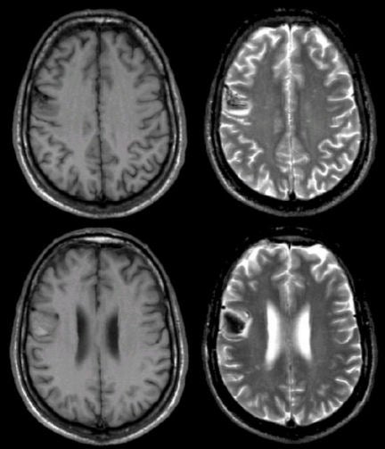
| Acute Intracerebral Hemorrhage: (Left
top and bottom) T1-weighted
axial MRIs; (Right top and bottom) T2-weighted axial MRIs. Note that
on the T1-weighted images, there
is an abnormality in the right posterior frontal lobe near the surface that is mostly isointense / hypointense. The same
area on the T2-weighted images is dark with a surrounding bright signal.
This is the characteristic picture of a hyperacute hemorrhage changing to an acute (approximately 3 days old) hemorrhage on MRI. The dark signal on T1- and T2-weighted images represents deoxyhemoglobin whereas the isointense signal is oxyhemoglobin. The surrounding bright signal is vasogenic edema. In this case, the hemorrhage was due to hypertension. The findings of blood on MRI are complex and depend on timing. To learn more, review the powerpoint slide show, Blood on MRI: Time-dependent Changes. |
Revised
11/29/06.
Copyrighted 2006. David C Preston.