|
An 89 year-old man presented with confusion, difficulty with comprehension, and right sided weakness affecting the leg more than the arm. |
![]()
![]()
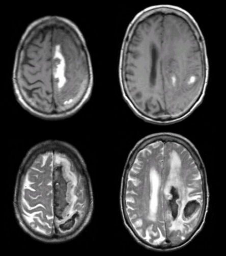
| Lobar Intracerebral Hemorrhage from Amyloid Angiopathy:
(Top) T1-weighted axial MRIs; (Bottom) T2-weighted axial MRIs. Note the large subacute hemorrhages.
One is in the left medial frontal lobe and the other is
in the left subcortical frontoparietal area. The hemorrhage is bright on
T1-weighted and dark on T2-weighted images, denoting intracellular methemoglobin. On T2-weighted
images,
there is also prominent surrounding vasogenic edema. In this case,
the hemorrhage was due to amyloid. Amyloid associated intracerebral
hemorrhage is common in very elderly individuals and often occurs as
a lobar hemorrhage, though subcortical hemorrhage can also be seen. The characteristics of blood on MRI are complex and depend on timing. In this case, the black rim is hemosiderin; the white interior is methemoglobin and the surrounding white area is vasogenic edema. To learn more, review the powerpoint slide show, Blood on MRI: Time-dependent Changes. |
Revised
11/23/06.
Copyrighted 2006. David C Preston