|
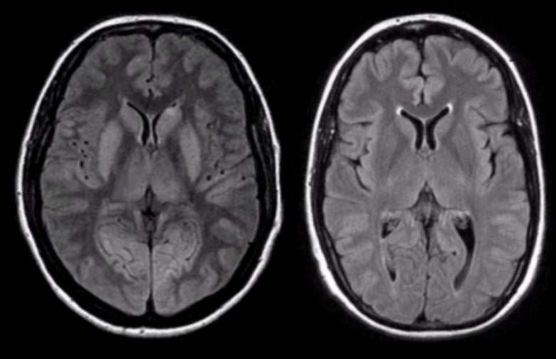
A 51 year-old man was unresponsive after a cardiac arrest. Neurological examination demonstrated intact brainstem reflexes and extensor posturing. No evidence of cortical function was present. |
![]()
![]()
| Anoxic Encephalopathy (Diffuse Cerebral Edema): Flair axial MRIs; (Left) patient - day 1; (Right) normal. Note that several abnormalities are present in the patient's scan, including: 1) an increased differentiation between grey and white matter; 2) effacement of the sulci; 3) abnormal signal in the basal ganglia; and 4) abnormal signal in the thalamus. Because the infarction is early, the increased differentiation between the gray and white matter is well seen, and is due to the brighter signal in the infarcted cortex and deep nuclei compared with the white matter. Note that later when the cerebral edema becomes more prominent, the differentiation between the gray and white matter will become decreased. The effacement of the sulci occurs from edema associated with the infarction. The bright signal in the basal ganglia and thalami in the Flair images results from infarction of these nuclei. This radiographic picture is most often seen following trauma, hypoxia (as in this case) or CNS infection, especially meningitis. In this scenario, the diffuse edema leads to increased intracranial pressure followed by decreased brain perfusion and subsequent brain death. |
Revised
11/29/06
Copyrighted 2006. David C Preston