|
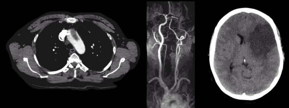
A 44 year-old woman presented with the acute onset of an expressive aphasia, a severe right hemiplegia, and left gaze deviation. She had systemic lupus erythematosus (SLE) associated with a lupus anticoagulant. |
![]()
![]()
![]()
| Aortic Embolus: (Left) CT scan of the chest with contrast. Note the large clot within the aortic arch; (Middle) Magnetic Resonance Angiogram - arch study. Note the complete cut-off of the left internal carotid artery, with the complete absence of other areas of vascular occlusion or plaque; (Right) Axial CT scan of the head. There is a large infarct involving the left basal ganglia and superior division of the left middle cerebral artery (MCA). This is the angiographic picture of an embolus. In this case, the patient was presumably hypercoagulable from her SLE, resulting in the thrombus in her aortic arch, which broke off and traveled proximally to occlude the internal carotid artery. |
Revised
11/29/06
Copyrighted 2006. David C Preston