![]()
![]()
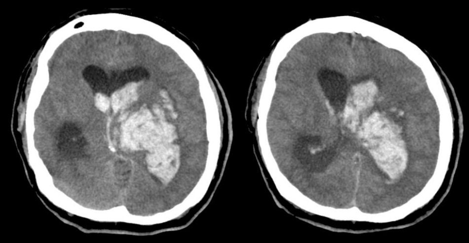
| Basal Ganglia and Thalamic Intracerebral Hemorrhage: Axial CT scans. Note the large intracerebral hemorrhage. The hemorrhage is so large that it involves the left basal ganglia and thalamus. Note that the blood has spread into the adjacent lateral ventricle. Also note that the lateral ventricles have become "trapped" because of compression of the third ventricle and the foramina of Monro. |
Revised
11/23/06.
Copyrighted 2006. David C Preston.