![]()
![]()
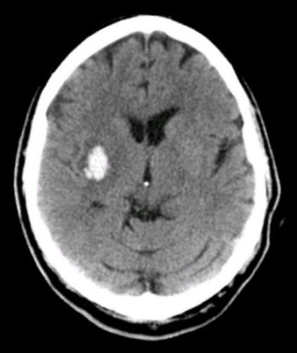
| Small Basal Ganglia (Putamen) Intracerebral Hemorrhage: Axial CT scan. Note the intracerebral hemorrhage originating in the area of the basal ganglia on the right. Before the advent of CT scans, intracerebral hemorrhages were thought to always be catastrophic neurological events. However, small hemorrhages can mimic ischemic strokes. This underscores the need to obtain a CT scan in every stroke patient to exclude the possibility of hemorrhage, especially before antiplatelet therapy or anticoagulation is considered. |
Revised
11/29/06.
Copyrighted 2006. David C Preston.