![]()
![]()
![]()
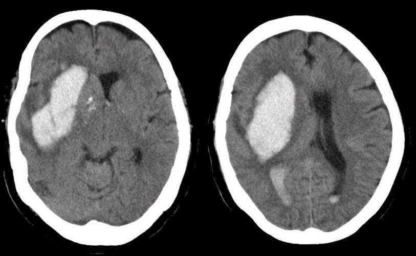
| Basal Ganglia (Putamen) Intracerebral Hemorrhage:
Axial CT scans. Note the large intracerebral hemorrhage originating in
the area of the basal ganglia on the right. If one looks closely at the occipital horns
of the lateral ventricles, one can appreciate blood (white signal) as well,
indicating that the hemorrhage has dissected into the ventricular system. The basal
ganglia is a common location of intracerebral bleeds due to hypertension. This is one of the common sites of hypertensive intracerebral hemorrhage. Hemorrhages in this location typically result in a contralateral hemiparesis affecting the face, arm and leg (from involvement of the internal capsule), associated with a hemisensory loss. With larger lesions, aphasia develops with lesions on the dominant side and neglect syndromes with lesions on the non-dominant side. With very large lesions, intracranial hypertension may develop, as manifested by headache, nausea and vomiting, and a depressed level of consciousness. |
Revised
11/15/06.
Copyrighted 2006. David C Preston.