|
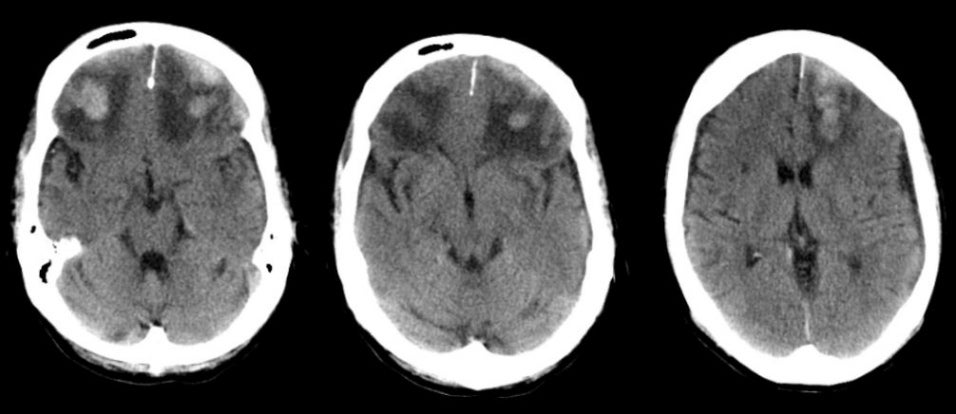
A 26 year-old man was brought to the hospital agitated and confused following a motor vehicle accident. |
![]()
![]()
| Frontal Pole Contusion. Axial CT scans without contrast. Note the areas of traumatic contusion, which consist of hemorrhage and surrounding edema, in both frontal lobes. There is also mass effect on the anterior horns of the lateral ventricles. The frontal poles are a common location for cerebral contusions following head injury, wherein the brain continues to move forward, striking the inner skull, after the head has stopped moving. |
Revised
11/15/06.
Copyrighted 2006. David C Preston