|
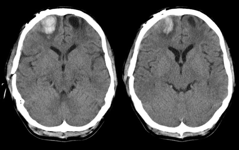
A 45 year-old man was brought to the hospital lethargic and not speaking following a motor vehicle accident. |
![]()
![]()
| Frontal Pole Contusion. Axial CT scans without contrast. Note the area of traumatic contusion, which consists of hemorrhage and surrounding edema, in the right frontal lobe. The low density area on the left is consistent with cerebral edema. The frontal poles are a common location for cerebral contusions following head injury, wherein the brain continues to move forward, striking the inner skull, after the head has stopped moving. |
Revised
11/15/06.
Copyrighted 2006. David C Preston