|
A 12 year-old girl presented with headaches which were worse with lying down or coughing. Her examination was notable for bilateral papilledema. |
![]()
![]()
![]()
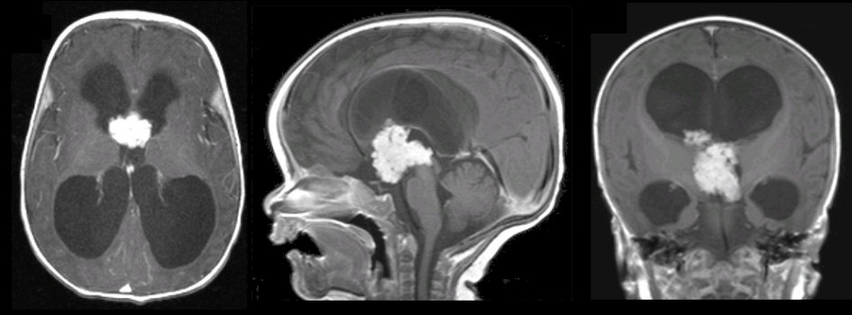
| Choroid Plexus Papilloma.
T1-weighted with gadolinium MRI scans; (Left) Axial; (Middle) Sagittal;
(Right) Coronal. Note the enhancing mass located in the third
ventricle and extending through the foramina of Monro into the
lateral ventricles. Also note the non-communicating hydrocephalus
with enlargement of the lateral and third ventricles, but not the
fourth ventricle. Surgical excision showed the tumor to be a choroid
plexus papilloma.
Cerebrospinal fluid (CSF) is produced by the choroid plexus within the ventricles. Choroid plexus papillomas are typically histologically benign tumors that arise from the choroid plexus. Patients typically present with signs and symptoms of hydrocephalus. The hydrocephalus can occur from two different mechanisms: the tumor can secrete increased amounts of CSF or the tumor can grow and obstruct the ventricular system (as in this case). |
Revised
11/25/06.
Copyrighted 2006. David C Preston