| Computed Tomography (CT) of the Brain: Basics | |
|
Computerized Tomography (CT) is one of the most commonly used tests in neurology and neurosurgery. CT revolutionized neurology and neurosurgery in the 1970s enabling visualization of intracranial structures with excellent resolution. Although MRI has supplanted CT in the evaluation of many neurological conditions, it still plays an important role, especially in the acute evaluation of stroke and head trauma patients. |
|
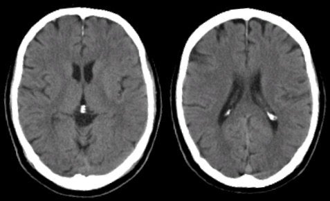
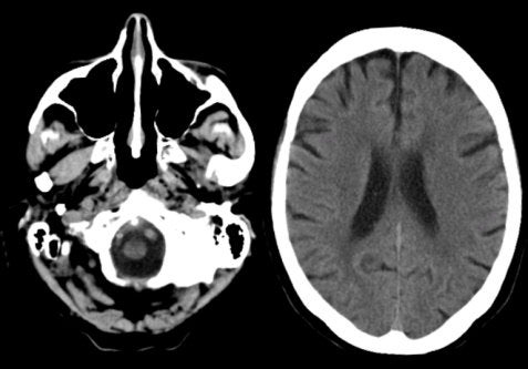
PHYSICS OF CT X-rays are applied in a circular motion with detectors on the opposite side of the body. Body tissue slices (typically 1 cm) are mathematically reconstructed and displayed on a gray scale matrix. The density of the tissue is in proportion to the attenuation of the x-rays which pass through. Tissues like air and water have little attenuation and are displayed as low densities (dark), whereas bone has high attenuation and is displayed as high density (bright) on CT. Among pathologic conditions, high density lesions are often seen with freshly clotted blood, hyperemia and with the use of contrast. Low density lesions include edema and necrosis. Similar to gadolinium in MRI, iodinated contrast agents are used in CT to demonstrate vascular structures and breakdown of the blood-brain barrier. The latter most often occur in tumors, infection and inflammation. |
|
NEUROLOGICAL INDICATIONS FOR CRANIAL CT CT is most commonly indicated in the emergency room evaluation of patients with acute head trauma and acute neurological dysfunction, primarily to look for suspected intracranial or subarachnoid hemorrhage. Its extremely rapid acquisition time and sensitivity of detecting hemorrhage makes it the modality of choice in the acute setting. CT is readily available at nearly all institutions. Claustrophobia is not a major issue, as it is in MRI. In general, CT is useful in the following conditions: ● Vascular - Ischemic stroke (> 2 days old) - Hemorrhagic stroke (acutely) ● Trauma ● Hydrocephalus With the use of contrast, it may also be helpful in the following conditions: ● Tumor (both primary CNS and metastatic) ● Infection and Abscess |
|
|
Show What is High Density (Bright) on CT in a Normal Patient |
|
|
|
 |
|
Show What is Low Density (Dark) on CT in a Normal Patient |
|
|
|
 |
LIMITATIONS OF CT • Subject to motion artifact • Artifacts from bone may interfere with detection of disease at the skull base and in the posterior fossa • Limited to axial views • Unable to detect acute ischemic stroke (first 1-2 days) • Unable to detect demyelination • Poorer spatial resolution compared to MRI • Often does not detect lacunar strokes, especially in the brainstem • Herpes encephalitis poorly visualized
compared to MRI |
|
| Important Note: On CT images, the slices are at an angle. Thus, structures near the top of the slice are more rostral than structures near the bottom of the slice. Note, this is different from MRI which has true axial slices. |
 |
CONTRAINDICATIONS TO CT There are few contraindications to CT. Relative contraindications to CT include: • Pregnancy (potential harm of ionizing radiation to the fetus) • Use of intravenous contrast agents is associated with infrequent but substantial risks: - Rare cases of anaphylaxis - Renal dysfunction (baseline creatinine required before contrast is given) |
|