|
A 66 year-old man presented with falling and a slowed gait. |
![]()
![]()
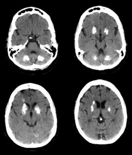
| Basal Ganglia and Cerebellar Calcifications:
Axial CT scans. Note the prominent calcifications in the
bilateral basal ganglia and cerebellum. This pattern of
calcifications can be
idiopathic (so-called Fahr's Disease), or as a secondary
phenomenon from hypoparathyroidism. Most often, these calcifications
are an incidental finding with no clinical significance. However,
in Fahr's Disease, patients may have deterioration of both motor and mental
functions. In addition, chorea and athetosis may be seen as late
phenomena. The etiology of this disorder is unknown; both sporadic
and familial cases have been reported. |
Revised
11/23/06.
Copyrighted 2006. David C Preston