|
A 63 year-old woman with known atrial fibrillation presented with acute left sided weakness. |
![]()
![]()
![]()
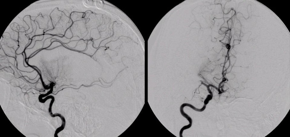
| Cardiac Embolus: Cerebral Angiogram, Right Carotid injection; (Left) Lateral view; (Right) AP view. Note the complete absence of any branches off the right middle cerebral artery (MCA) and the abrupt cutoff of the distal stem of the right MCA. The lenticulostriate vessels are particularly well seen. On the AP view, the right middle cerebral artery is abruptly occluded. This is the angiographic picture of an embolus, in this case of cardiac origin from the atrial fibrillation. In the anterior circulation, emboli commonly go to the middle, posterior and anterior cerebral arteries. In the posterior circulation, they typically go to the top of the basilar or the posterior cerebral arteries. (ACA = anterior cerebral artery; ICA = internal carotid artery). |
Revised
11/07/06
Copyrighted 2006. David C Preston