|
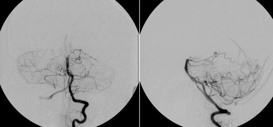
A 56 year-old man presented with chest pain and shortness of breath. EKG and cardiac enzymes were consistent with an acute myocardial infarction. Four hours later, he abruptly went into a coma. |
![]()
![]()
![]()
| Cardiac Embolus: Cerebral Angiogram, Left Vertebral injection; (Left) AP view; (Right) Lateral view. Note the abrupt cutoff at the top of the basilar artery. There is the complete absence of any branches off the posterior cerebral arteries (PCAs). This is the angiographic picture of an embolus, in this case of cardiac origin. In this case, the MI and presumed wall motion abnormalities resulted in clot formation and the source of the embolus. In the anterior circulation, emboli commonly go to the middle, posterior and anterior cerebral arteries. In the posterior circulation, they typically go to the top of the basilar or the posterior cerebral arteries. (SCA = superior cerebellar artery) |
Revised
11/30/06
Copyrighted 2006. David C Preston