|
A 56 year-old man with known mitral stenosis and atrial fibrillation presented with confusion, cortical blindness and impaired vertical gaze. |
![]()
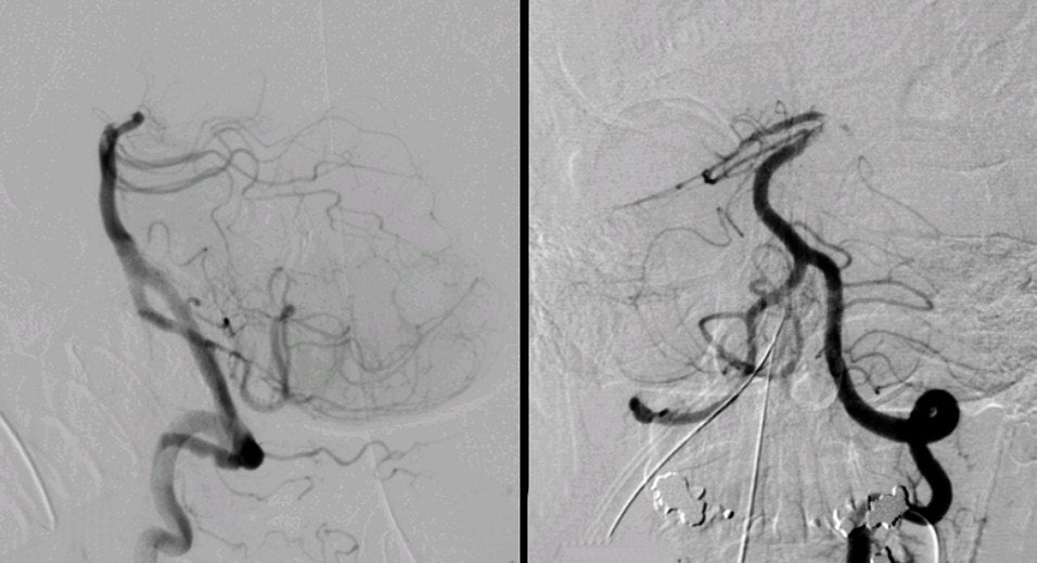
| Cardiac Embolus: Cerebral Angiogram, Left Vertebral injection; (Left) Lateral view; (Right) AP view. Note the abrupt cutoff at the top of the basilar artery. There is a nearly complete absence of any branches off the posterior cerebral arteries. This is the angiographic picture of an embolus, in this case of cardiac origin from the mitral stenosis and atrial fibrillation. In the anterior circulation, emboli commonly go to the middle, posterior and anterior cerebral arteries. In the posterior circulation, they typically go to the top of the basilar or the posterior cerebral arteries. (AICA = anterior inferior cerebellar artery, PCA = posterior cerebral artery, PICA = posterior inferior cerebellar artery, SCA = superior cerebellar artery). |
Revised
11/22/06
Copyrighted 2006. David C Preston