|
A 24 year-old woman presented with right neck pain and a right Horner's syndrome after a minor motor vehicle accident. Otherwise her neurological examination was normal. |
![]()
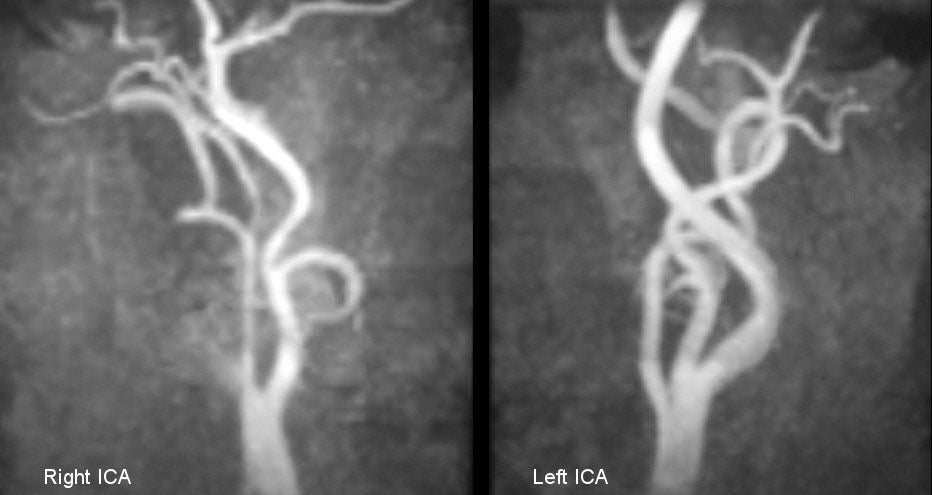
| Carotid Artery Dissection:
Magnetic Resonance Angiogram of the neck. Note the "string sign" of
the right internal carotid artery (ICA), compared to the normal image of the left
internal carotid artery. Although not specific for dissection,
this pattern is highly suggestive. If
the diagnosis of dissection is suspected, fat-suppression MRI is the
imaging study of choice. Intramural blood can be well demonstrated
on these scans. Routine magnetic resonance angiography (MRA) will
often demonstrate narrowing or occlusion of the vessel, but in most
cases cannot differentiate dissection from other etiologies. It is
critical to detect a dissection before ischemia occurs, so that
treatment can be promptly initiated. Arterial dissection occurs due to a tear in the intimal layer of the artery. The tear allows blood to enter the wall and form an intramural hematoma. Depending on which layer of the blood vessel is involved, either a subintimal or a subadventitial hematoma develops. A subintimal hematoma tends to cause stenosis of the artery, whereas a subadventitial hematoma often results in aneurysmal dilatation of the artery. In the case of stenosis, sluggish blood flow distal to the dissection results in the formation of fibrin clot. The clot continues to enlarge and eventually breaks off to travel and dislodge downstream as an embolus. Although rare in the general population, dissections of the carotid and vertebral arteries account for a substantial number of strokes in young adults and middle-aged patients. Most dissections involve some type of trauma or stretch to the head or neck. Sometimes, the trauma is trivial and forgotten by the patient. There is a higher incidence in certain congenital connective tissue disorders, including Marfan's syndrome, cystic medial necrosis, and fibromuscular dysplasia. The recognition of an arterial dissection requires a high degree of clinical suspicion and an ability to recognize some key neurological signs which help to key in on the diagnosis. The classic symptoms and signs of carotid artery dissection include the following: • Pain or headache on one side of the
head, face or neck |
Revised
11/30/06
Copyrighted 2006. David C Preston