|
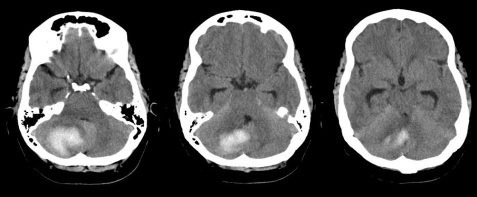
A 47 year-old hypertensive woman presented with headache, nausea and vomiting. On exam, she was unable to walk. |
![]()
![]()
![]()
| Cerebellar Intracerebral Hemorrhage:
Axial CT scans.
Note the area of high signal intensity in the right cerebellum which
denotes acute blood on CT imaging. Also note the effacement of the
adjacent fourth ventricle and the presence of early hydrocephalus
(enlargement of the temporal horns).
The cerebellum is one of the common locations for intracerebral hypertensive hemorrhage. If the hematoma expands, it can result in direct compression of the brainstem, or compression and obstruction of the fourth ventricle and secondary hydrocephalus (as seen above). It is relatively uncommon for patients with a cerebellar hemorrhage to display limb ataxia of the arms or legs (classic cerebellar signs). They are much more likely to present with nausea, vomiting and the inability to walk. Of all the possible locations for an intracerebral hemorrhage, the cerebellum is the most important to recognize early, as it is potentially treatable by surgical decompression and evacuation. If the hemorrhage results in compression of the fourth ventricle or aqueduct, acute hydrocephalus can develop, which is treatable by urgent shunting. |
Revised
11/30/06.
Copyrighted 2006. David C Preston