|
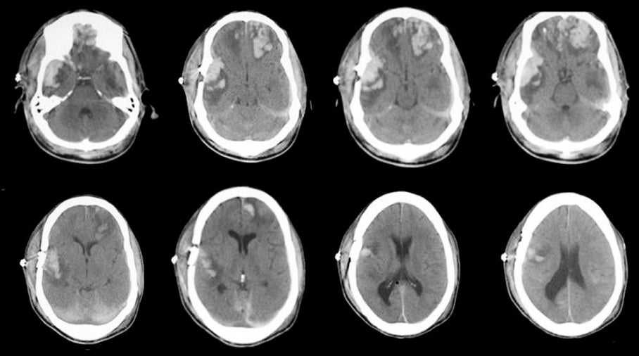
A 44 year-old woman was brought to the hospital in coma following a motor vehicle accident. |
![]()
![]()
![]()
![]()
| Multiple Contusions. Axial CT scans without contrast. Note the large areas of traumatic contusion, which consist of hemorrhage and surrounding edema, in the frontal lobes (left larger than right) and the right temporal lobe. Subarachnoid blood is also present, most easily seen over the tentorium posteriorly. A skull fracture is seen underlying the right temporal bone. The frontal and temporal lobe tips are common locations for cerebral contusions following head injury, wherein the brain continues to move forward, striking the inner skull, after the head has stopped moving. Contact is made first in the frontal and temporal tips. |
Revised
11/15/06.
Copyrighted 2006. David C Preston