|
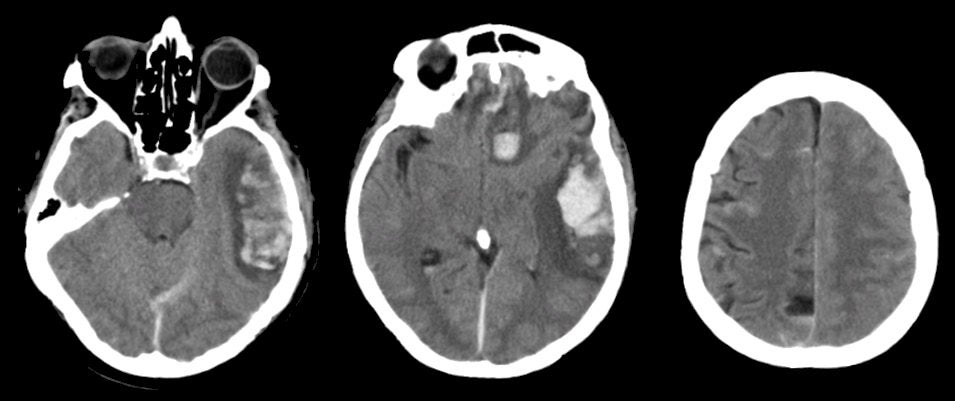
A 62 year-old woman was found lethargic at the bottom of a flight of stairs after a fall. |
![]()
![]()
![]()
| Multiple Contusions. Axial CT scans without contrast. Note the large areas of traumatic contusion, which consist of hemorrhage and surrounding edema, in the left inferior frontal lobe and the left temporal lobe. Subarachnoid blood is also present, seen in the interhemispheric fissure and in several of the sulci over the right convexity. The frontal and temporal lobe tips are common locations for cerebral contusions following head injury, wherein the brain continues to move forward, striking the inner skull, after the head has stopped moving. Contact is made first in the frontal and temporal tips. |
Revised
11/15/06.
Copyrighted 2006. David C Preston