|
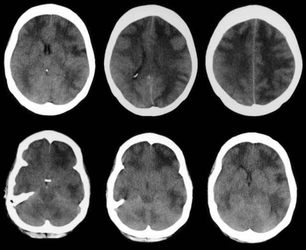
A 42 year-old man was drowsy and confused following a hypotensive episode. Within a day, his neurological condition markedly deteriorated and he fell into a coma. |
![]()
![]()
![]()
![]()
| Anoxic Encephalopathy (Diffuse Cerebral Edema): Axial CT scans, day 1. Note the diffuse cerebral edema, most prominently seen in the white matter. No sulci are seen over the convexities and the cisterns around the brainstem have been effaced. This radiographic picture is most often seen following trauma, hypoxia (as in this case) or CNS infection, especially meningitis. In this scenario, the diffuse edema leads to increased intracranial pressure followed by decreased brain perfusion and subsequent brain death. |
Revised
11/30/06
Copyrighted 2006. David C Preston