|
A 55 year-old woman presented with headaches followed by a series of strokes over 3 weeks. |
![]()
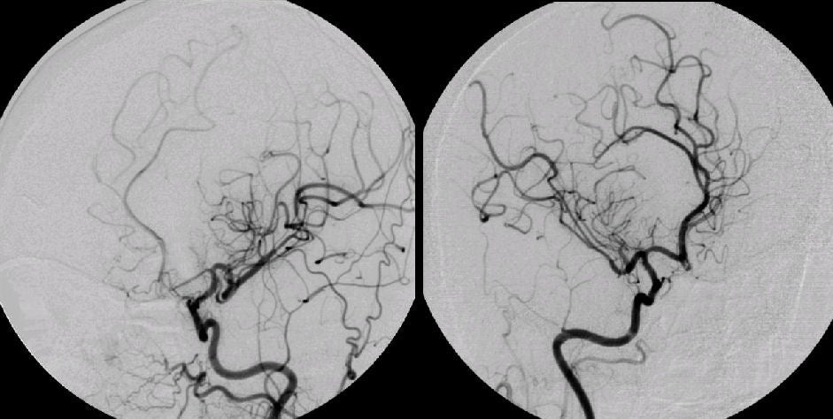
| Cerebral Vasculitis: Cerebral Angiogram, (Left) Left Internal Carotid Artery injection; (Right) Right Internal Carotid Artery injection; both oblique views. Note the severe tapered narrowing of the left proximal anterior cerebral artery. This appearance is consistent with cerebral vasculitis, which can occur as an isolated syndrome or as part of a more widespread systemic vasculitis. A similar appearance may occur from vasospasm following subarachnoid hemorrhage. Often, brain and meningeal biopsy are needed to confirm the diagnosis. Cerebral vasculitis often presents as an encephalopathy with superimposed focal deficits from multiple ischemic strokes. The syndrome can be confused with embolic infarctions and atherosclerotic intracranial disease. The latter is more prominent in individuals of Asian and African American descent compared to Caucasians. ICA = internal carotid artery; MCA = middle cerebral artery; ACA = anterior cerebral artery. |
Revised
11/22/06
Copyrighted 2006. David C Preston