|
A 33 year-old man presented with vertical nystagmus. Otherwise, his examination was normal. |
![]()
![]()
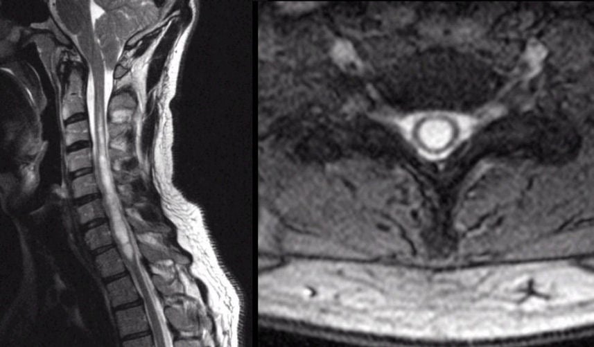
| Chiari Malformation Type I with Syrinx: (Left) T2-weighted
sagittal MRI of the spine; (Right)
T2-weighted axial image at the level of C5. Note
the Chiari type I malformation associated with the cervical syrinx,
wherein the brainstem and cerebellar tonsils descend
below the foramen magnum. Note the large syrinx in the cervical cord. On the axial scan,
one can see how large the syrinx is
compared to the spinal cord. In this case, the syrinx was completely
asymptomatic for many years, underscoring that a very chronic lesion can be
well compensated for. This malformation is know as the Chiari malformation, type I. There are several types of Chiari malformations. In Type I, the malformation is limited to descent of the medulla and cerebellum into the spinal canal. Some cases are associated with syringomyelia and/or syringobulbia. This malformation is often asymptomatic. When symptomatic, patients typically develop posterior headaches and dizziness. In more severe cases, patients may develop vertical nystagmus and other cranial nerve abnormalities. |
Revised
11/23/06
Copyrighted 2006. David C Preston