|
A 48 year-old woman presented with intermittent headaches for several weeks followed by a continuous severe headache associated with vomiting. |
![]()
![]()
![]()
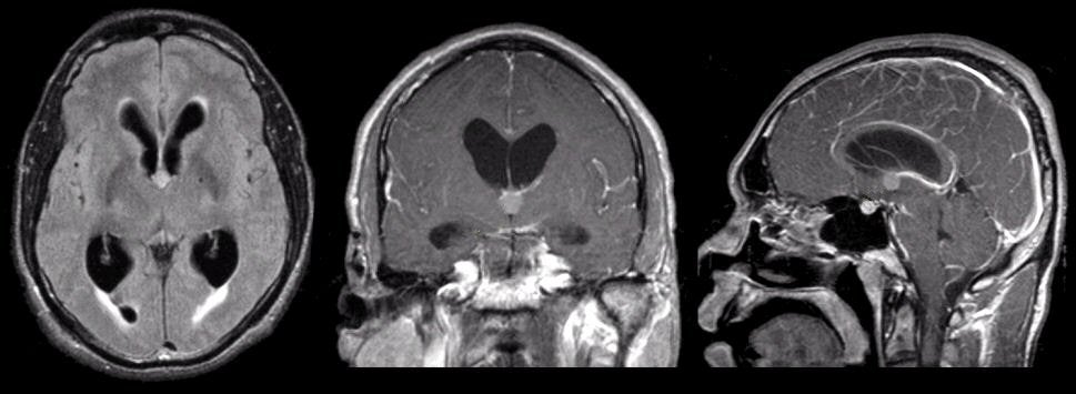
| Colloid Cyst of the Third Ventricle:
(Left) Flair axial MRI; (Middle) T1-weighted
with gadolinium coronal MRI; (Right) T1-weighted with gadolinium sagittal MRI. Note the
cyst in the anterior third ventricle which is obstructing both
foramina of Monro, causing hydrocephalus of the lateral ventricles. On the Flair
image, the transependymal edema can be seen capping the anterior and
posterior horns of the lateral ventricles. This lesion is a colloid cyst, which is a benign congenital cyst that arises in the anterior third ventricle. They are often asymptomatic. However, if the cyst enlarges, compressing the foramina of Monro, non-communicating hydrocephalus may develop. Classically, this results first in intermittent severe headaches due to intermittent obstruction of the foramina of Monro. Rare cases result in sudden death. |
Revised
11/04/06
Copyrighted 2006. David C Preston