| A 51 year-old man presented with severe nausea. While in the hospital, he was noted to have episodes of shaking of the arms and would not respond to verbal stimuli. An urgent head CT scan was ordered, followed by emergency neurosurgical intervention and a subsequent MRI scan. |
![]()
![]()
![]()
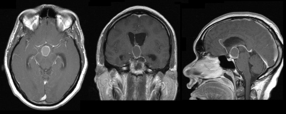
| Craniopharyngioma.
(Left) T1-weighted with
gadolinium axial MRI; (Middle) T1-weighted with gadolinium coronal
MRI; (Right) T1-weighted with gadolinium sagittal MRI;. Note the presence of a large suprasellar
mass that enhances with gadolinium. If one looks closely, one can
see the
thickening and greater enhancement of the mass near the infundibulum
of the pituitary. Surgical removal
demonstrated a craniopharyngioma. Craniopharyngiomas arise in the
suprasellar region and are often calcified and cystic. They are slow
growing tumors that occur in children and adults, and can become
very large in size. In children, they are thought to occur as a
result of impaired embryogenesis of structures in or near the
infundibulum of the pituitary gland. In adults, they are believed to
occur as a result of metaplasia of pituitary squamous epithelium.
Similar to pituitary macroadenomas, they may present with endocrine
dysfunction or focal neurological signs due to mass effect in the
suprasellar region. |
Revised
11/28/06.
Copyrighted 2006. David C Preston