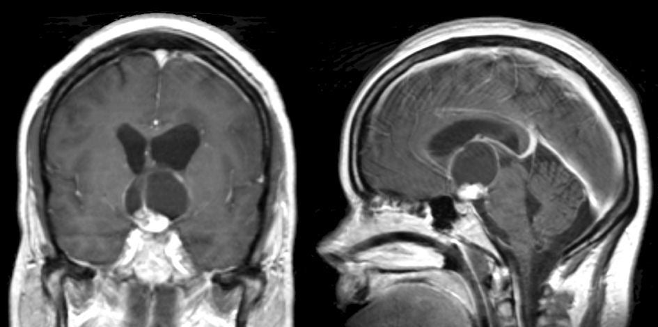
| A 32 year-old man presented with progressive headaches and trouble with his vision. |
![]()
![]()
![]()
| Craniopharyngioma. (Left) T1-weighted with gadolinium
coronal MRI; (Right) T1-weighted with gadolinium sagittal MRI. Note
the presence of a suprasellar mass that enhances with gadolinium and
is associated with a large cystic component. Also note that the
cystic part of the tumor obstructs the foramen of Monro, worse
on the left compared to the right side. Surgical removal demonstrated
a craniopharyngioma. Craniopharyngiomas arise in the suprasellar
region and are often calcified and cystic. They are slow growing
tumors that occur in children and adults, and can become very large
in size. In children, they are thought to occur as a result of
impaired embryogenesis of structures in or near the infundibulum of
the pituitary gland. In adults, they are believed to occur as a
result of metaplasia of pituitary squamous epithelium. Similar to
pituitary macroadenomas, they may present with endocrine dysfunction
or focal neurological signs due to mass effect in the suprasellar
region. |
Revised
11/29/06.
Copyrighted 2006. David C Preston