|
A 33 year-old woman presented with complex partial seizures which often generalized. On examination, there was subtle right sided weakness. |
![]()
![]()
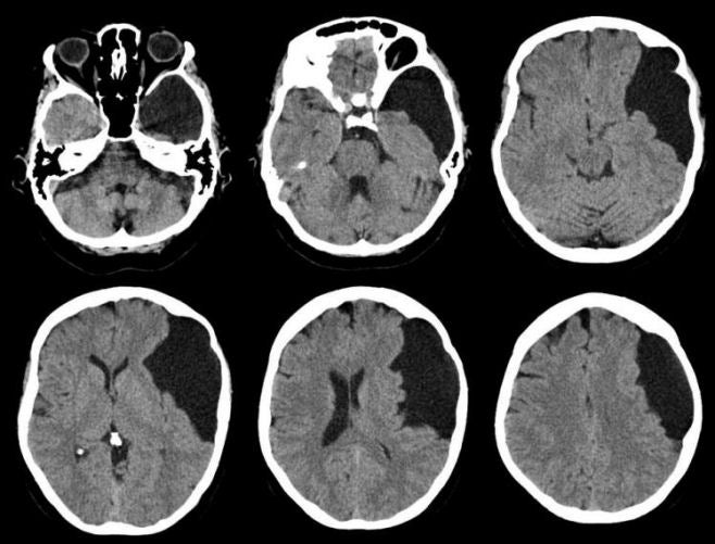
| Arachnoid Cyst of the Middle Cranial Fossa:
Axial CT scans. Note
the large, left sided cystic lesion.
Also, note the mild mass effect on the left hemisphere (i.e. compression of the
left frontal lobe and lateral ventricle, with a slight shift of the pineal gland from left to right). The skull immediately adjacent to the cyst
is thinned. Arachnoid cysts are common congenital malformations. They contain fluid but generally do not communicate with the ventricular system. The most common locations are the Sylvian fissure and middle cranial fossa (as in the case above), suprasellar cistern, quadrigeminal cistern, cerebellopontine angle, posterior infratentorial midline, cerebral convexity and interhemispheric fissure. Most are asymptomatic and found incidentally on brain imaging studies. Rarely, they can enlarge and present as a mass lesion with focal neurological signs, seizures or hydrocephalus. |
Revised
11/19/06
Copyrighted 2006. David C Preston