|
A 22 year-old woman presented for evaluation of headaches. On examination, there were subtle neurological signs suggestive of mild right sided weakness. |
![]()
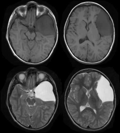
| Arachnoid Cyst of the Middle Cranial Fossa:
(Top)
T1-weighted axial MRIs; (Bottom) T2-weighted axial MRIs. Note
the large cyst in the left middle cranial fossa. It has replaced most of the
anterior temporal lobe as well as much of the posterior frontal lobe.
The cyst contains fluid; thus it is dark on T1-weighted and bright on
T2-weighted images. Arachnoid cysts are common congenital malformations. They contain fluid but generally do not communicate with the ventricular system. The most common locations are the Sylvian fissure and middle cranial fossa (as in the case above), suprasellar cistern, quadrigeminal cistern, cerebellopontine angle, posterior infratentorial midline, cerebral convexity and interhemispheric fissure. Most are asymptomatic and found incidentally on brain imaging studies. Rarely, they can enlarge and present as a mass lesion with focal neurological signs, seizures or hydrocephalus. |
Revised
11/29/06
Copyrighted 2006. David C Preston