|
A 47 year-old man developed mid-back pain followed by numbness and weakness of both legs over the next two weeks, associated with urinary incontinence. He had been treated for Staphylococcus aureus pyelonephritis three weeks earlier. |
![]()
![]()
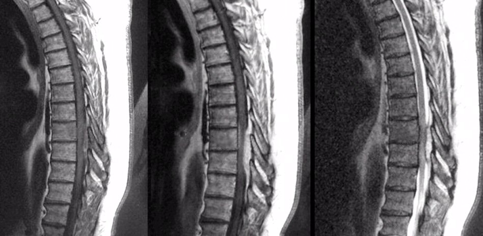
| Epidural Abscess: Sagittal MRIs of the
Thoracic Spine; (Left) T1-weighted; (Middle) T1-weighted with gadolinium; (Right) T2-weighted. Note the
lesion which is bright on T2-weighted images and enhances with gadolinium (middle
image compared
to the left). The lesion is clearly extra-axial and compresses the spinal
cord. Emergency surgery demonstrated an epidural abscess with gram positive cocci. Epidural abscesses most often occur from spread through the blood system to the vertebral body, directly to the epidural space, or both. Patients most often present subacutely over days with fever and spine pain, often followed by radicular symptoms and then frank spinal cord compromise. |
Revised
11/30/06
Copyrighted 2006. David C Preston