|
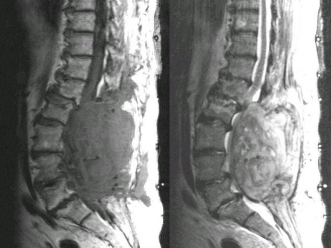
A 47 year-old man underwent routine lumbar laminectomy for a herniated disk. Following the surgery, he developed progressive leg weakness and then paraplegia and urinary incontinence. On examination, he had a sensory level at L1, paraplegia, and no reflexes in the legs. |
![]()
![]()
| Spinal Epidural Hematoma: MRI of the Thoracic Spine; (Left) T1-weighted; (Right) T2-weighted. Note the large extradural lesion which compresses the thecal sac from L2 through S1. It is isointense on the T1-weighted scan and slightly bright on the T2-weighted scan. This is a an acute epidural hematoma. The patient subsequently underwent emergency decompressive surgery. |
Revised
11/22/06
Copyrighted 2006. David C Preston