|
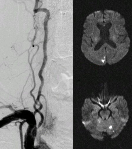
A 73 year-old man presented with nausea, vomiting and an unsteady gait. On examination, he was noted to have a left visual field defect. |
![]()
![]()
| Extracranial Vertebral Stenosis: (Left) Angiogram of the right subclavian artery. Note the high grade stenosis at the origin of the vertebral artery. (Right) Diffusion-weighted MRI. Note the areas of acute infarction in the rostral cerebellum on the left, and the right occipital lobe. These lesions were the cause of the patient's symptoms. The vertebral stenosis was successfully treated by placement of a stent. |
Revised
11/30/06
Copyrighted 2006. David C Preston