|
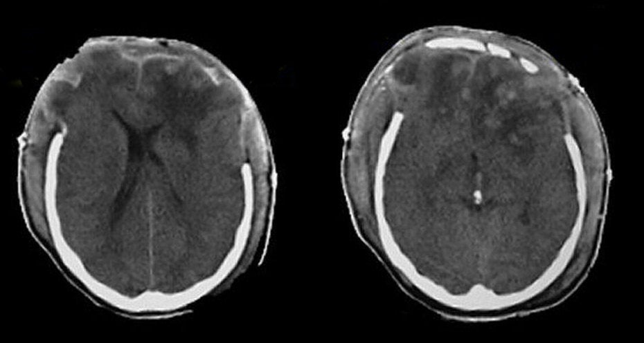
A 35 year-old man suffered a closed head injury during a sports activity. Bilateral craniectomies were done emergently for massive bilateral subdural and epidural hematomas. Post-operatively, he remained stuporous and a repeat CT scan was performed. |
![]()
| Brain Herniation Through Craniectomy Sites:
Axial CT scans. Note the areas of edema and
contusion in both frontal lobes. Also note the herniation of brain tissue through the craniectomy sites. In this case, the craniectomy defects allowed
a path of herniation for the brain tissue and potentially avoided transtentorial herniation. The displacement of brain structures described above illustrates the Monro-Kellie doctrine, which states that in an adult the cranial volume is a constant. The cranial contents consist primarily of brain, cerebrospinal fluid (CSF) and blood vessels. If a mass such as a hematoma, tumor or edema develops, these elements must shift to accommodate the mass. Since the cranial volume is a constant, part of the cranial contents will herniate to make room for the mass. In this case, the expansion of the brain from swelling resulted in herniation of brain tissue out of the skull through the surgical defects. |
Revised
11/18/06.
Copyrighted 2006. David C Preston