|
A 25 year-old woman developed progressive confusion and lethargy over six weeks. |
![]()
![]()
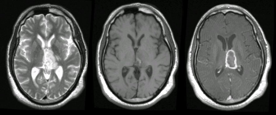
| Glioblastoma Multiforme (Thalamus).
(Left) T2-weighted axial MRI;
(Middle) T1-weighted axial MRI; (Right) T1-weighted with gadolinium axial MRI. Note
the mass in the left
thalamus that ring enhances with
gadolinium (GAD).
Also note the mass effect on the adjacent third ventricle, seen best
on the T1-weighted image. Stereotactic biopsy showed glioblastoma multiforme.
Glioblastoma multiforme (GBM), also referred to as a Grade IV
astrocytoma, is the most common type of primary brain tumor. It is a
malignant tumor that carries a very poor prognosis, and typically
results in death in 2 years. On CT and MRI imaging, the tumor is
often large, irregular and infiltrative, and located in the white
matter with surrounding edema. Histologically, the tumor is highly
cellular and anaplastic with necrosis. Associated hemorrhage is not
uncommon. Clinically, patients present with slowly progressive focal neurological signs, and signs of increased intracranial pressure (i.e., headache, nausea, and vomiting). Seizures may be an initial presentation or may occur later in the course. |
Revised
11/25/06.
Copyrighted 2006. David C Preston