|
A 49 year-old woman with secondary progressive multiple sclerosis (MS) presented with confusion and right sided weakness over 2 weeks. |
![]()
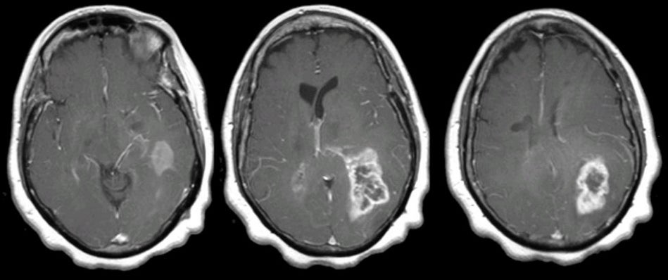
| Glioblastoma Multiforme (Parietal Lobe).
T1-weighted with gadolinium axial MRIs. Note the large
enhancing mass in the left posterior parietal area that demonstrates mass effect. This picture is most suggestive of an intrinsic CNS malignancy such as glioblastoma multiforme. Biopsy confirmed the diagnosis
of malignant glioblastoma. Glioblastoma multiforme (GBM), also
referred to as a Grade IV astrocytoma, is the most common type of
primary brain tumor. It is a malignant tumor that carries a very
poor prognosis, and typically results in death in 2 years. On CT and
MRI imaging, the tumor is often large, irregular and infiltrative,
and located in the white matter with surrounding edema.
Histologically, the tumor is highly cellular and anaplastic with
necrosis. Associated hemorrhage is not uncommon. Clinically, patients present with slowly progressive focal neurological signs, and signs of increased intracranial pressure (i.e., headache, nausea, and vomiting). Seizures may be an initial presentation or may occur later in the course. |
Revised
11/25/06.
Copyrighted 2006. David C Preston