|
A 43 year-old man developed neck pain radiating to both shoulders. Gait was stiff and awkward. Examination was notable for increased reflexes in the arms and legs, and bilateral Babinski responses. |
![]()
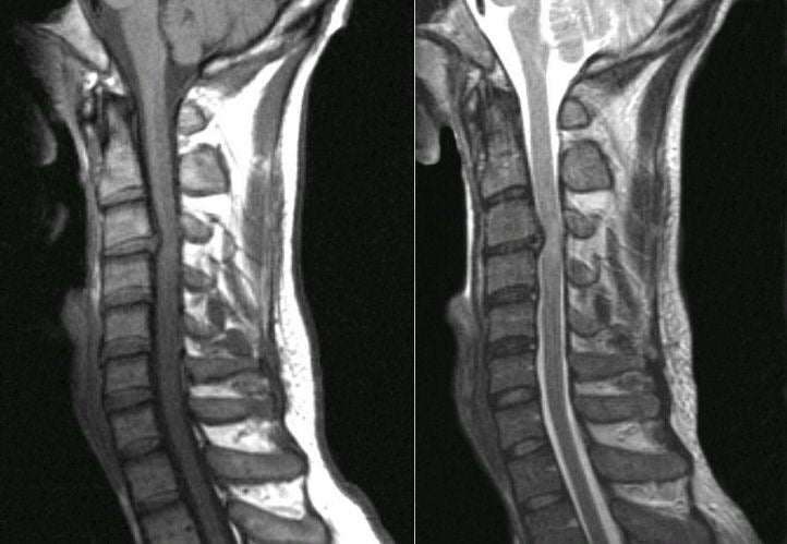
| Herniated C3-4 Disk:
Sagittal MRI scans of
the cervical spine; (Left) T1-weighted; (Right) T2-weighted. Note
the large disk herniation between the C3 and C4 vertebral bodies, deforming the
spinal cord at that level. Also note the effacement of the CSF both anteriorly
and posteriorly, best seen on the T2-weighted scan (where CSF is bright). In
this case, the spinal cord compression resulted in the increased
reflexes in the upper and lower extremities, and bilateral Babinski
responses. Among spinal disorders, herniated disks are very common. Disks are located between the vertebral bodies. They are composed of a thick annulus fibrosis surrounding a jelly like interior known as the nucleus pulposus. The anterior and posterior longitudinal ligaments run along the front and back of the vertebral bones and disks respectively. Disk herniations most often occur posterior laterally, where they compress the exiting nerve root at that level. Straight posterior disk herniations, although less common, may result in direct spinal cord or cauda equina compression. |
Revised
11/30/06
Copyrighted 2006. David C Preston