|
A 29 year-old man developed low back pain radiating to the right anterior thigh. Examination showed an absent right knee reflex, weakness of right hip adduction and knee extension, and loss of sensation over the right anterior thigh and medial calf. |
![]()
![]()
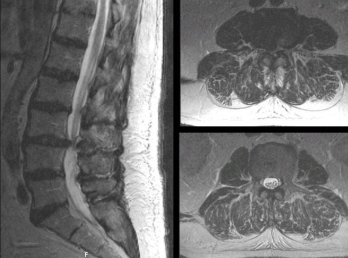
| Herniated L3-4 Disk:
(Left) T2-weighted sagittal MRI of
the lumbosacral spine; (Right Upper) T2-weighted axial MRI at the L3-4 level; (Right
Lower) T2-weighted axial MRI at the L5 level. On the T2-weighted images, CSF is bright. Note
the large disk herniation between the L3 and L4 vertebral bodies. On the axial scans, the
disk herniation
essentially obliterates the CSF (right upper scan). Compare this scan to
the level just below the compression (right
lower scan), where there is abundant CSF. If this herniation were to enlarge, this patient would be at risk for
developing a cauda equina syndrome. Among spinal disorders, herniated disks are very common. Disks are located between the vertebral bodies. They are composed of a thick annulus fibrosis surrounding a jelly like interior known as the nucleus pulposus. The anterior and posterior longitudinal ligaments run along the front and back of the vertebral bones and disks respectively. Disk herniations most often occur posterior laterally, where they compress the exiting nerve root at that level. Straight posterior disk herniations, although less common, may result in direct spinal cord or cauda equina compression. |
Revised
11/30/06
Copyrighted 2006. David C Preston