|
A 39 year-old man developed low back pain radiating down the right leg associated with numbness of the lateral foot. Examination showed an absent right ankle reflex, weakness of right knee and ankle plantar flexion, and patchy loss of sensation over the right lateral foot and posterior lateral leg. |
![]()
![]()
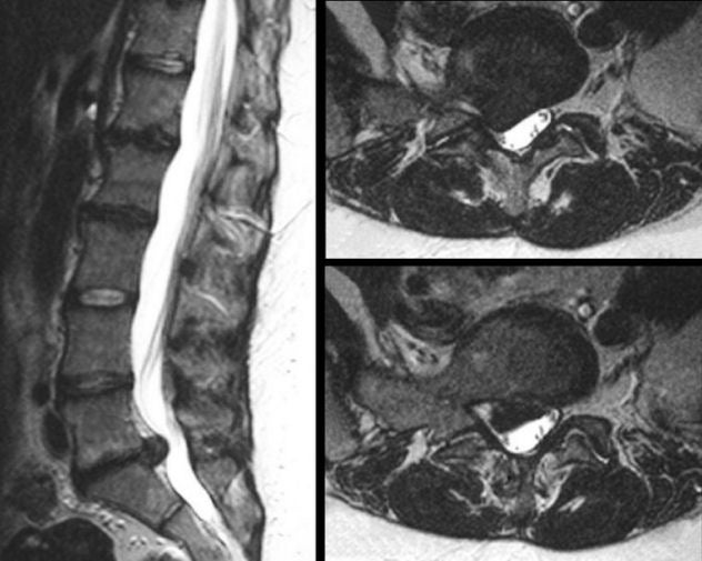
| Herniated L5-S1 Disk:
T2-weighted MRI scans of
the lumbosacral spine; (Left) Sagittal image; (Right) Axial images. On the T2-weighted images, CSF is bright. Note
the large disk herniation between the L5 and S1 vertebral bodies. On the axial scans, the herniation is posterior lateral, compressing the exiting S1 nerve root,
resulting in the clinical symptoms and signs of an S1 radiculopathy. Note
how the right foramen is nearly completed occluded.
Among spinal disorders, herniated disks are very common. Disks are located between the vertebral bodies. They are composed of a thick annulus fibrosis surrounding a jelly like interior known as the nucleus pulposus. The anterior and posterior longitudinal ligaments run along the front and back of the vertebral bones and disks respectively. Disk herniations most often occur posterior laterally, where they compress the exiting nerve root at that level. Straight posterior disk herniations, although less common, may result in direct spinal cord or cauda equina compression. |
Revised
11/30/06
Copyrighted 2006. David C Preston