|
A 33 year-old man developed severe low back pain radiating down the left leg. Examination showed an absent left ankle reflex and loss of sensation over the left lateral foot and posterior lateral leg. Straight leg raise on the left caused severe pain radiating down from the buttock to the ankle. |
![]()
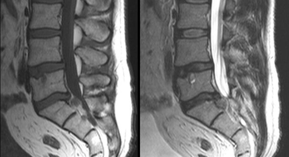
| Herniated L5-S1 Disk:
(Left) T1-weighted sagittal MRI; (Right) T2-weighted sagittal MRI.
Note the large disk herniation between the L5 and S1 vertebral
bodies. If one looks closely at the T1-weighted scan (left),
one can see that a fragment has actually separated from the disk. This is known as a
free fragment. In such cases, the fragment may stay at the same
level or migrate rostrally or caudally. Among spinal disorders, herniated disks are very common. Disks are located between the vertebral bodies. They are composed of a thick annulus fibrosis surrounding a jelly like interior known as the nucleus pulposus. The anterior and posterior longitudinal ligaments run along the front and back of the vertebral bones and disks respectively. Disk herniations most often occur posterior laterally, where they compress the exiting nerve root at that level. Straight posterior disk herniations, although less common, may result in direct spinal cord or cauda equina compression. |
Revised
11/30/06
Copyrighted 2006. David C Preston