|
A 33 year-old man developed severe low back pain radiating down the left leg. Examination showed an absent left ankle reflex and loss of sensation over the left lateral foot and posterior lateral leg. Straight leg raise on the left caused severe pain radiating down from the buttock to the ankle. |
![]()
![]()
![]()
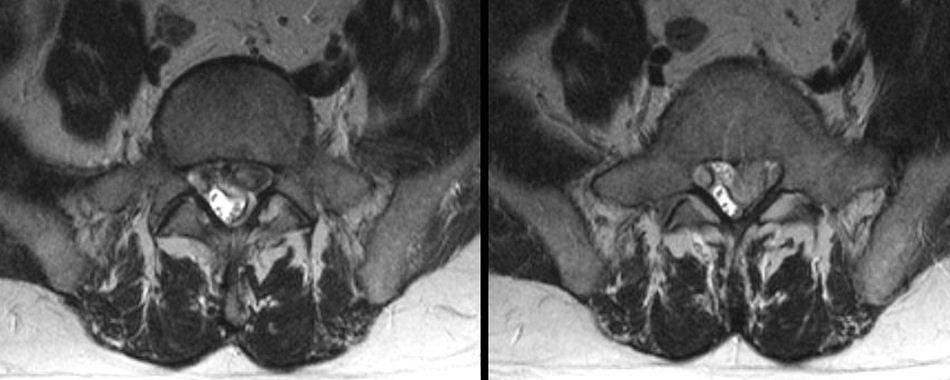
| Herniated L5-S1 Disk. (Left) T2-weighted axial MRI at the
L5-S1 level; (Right) T2-weighted axial MRI at the S1 vertebral body
level. Note the large disk herniation that has migrated from
the L5-S1 level caudally to the S1 vertebral level. Also note the
prominent compression and distortion of the thecal sac,
resulting in the clinical symptoms and signs of an S1 radiculopathy. Among spinal disorders, herniated disks are very common. Disks are located between the vertebral bodies. They are composed of a thick annulus fibrosus surrounding an jelly like interior known as the nucleus pulposus. Along the front and back of the vertebral bones and disks are the anterior and posterior longitudinal ligaments, respectively. Thus, disk herniations most often are posterior lateral where they compress the exiting nerve root at that level. Straight posterior disk herniations, although less common, may result in direct spinal cord or cauda equina compression. |
Revised
11/30/06
Copyrighted 2006. David C Preston