|
A 58 year-old woman developed right neck and shoulder pain followed by paresthesias down the right arm. Two weeks later, she noted her right foot was limping. On examination, her right biceps reflex was depressed. The right knee and ankle reflexes were pathologically brisk. |
![]()
![]()
![]()
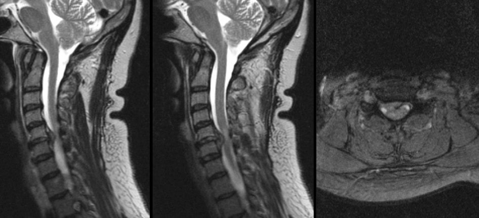
| Herniated C5-6 Disk:
(Left) T2-weighted right parasagittal MRI; (Middle) T2-weighted
mid-sagittal MRI; (Right) T2-weighted axial MRI at the level of
C5-6. Note the large disk herniation between the C5 and C6
vertebral bodies. On the axial scan, the disk herniation is
posterior lateral to the right, compressing the exiting C6 nerve
root. Also note the effacement of the CSF
and compression of the spinal cord. If one looks closely at the cord,
one can see bright signal
within the cord. This finding occurs either from gliosis or ischemia
from the compressing disk, and portends a poorer prognosis. In this
case, the spinal cord compression resulted in the brisk reflexes
and weakness in the right lower extremity,
while the nerve root compression resulted in the paresthesias down
the right arm and depressed right biceps reflex. Among spinal disorders, herniated disks are very common. Disks are located between the vertebral bodies. They are composed of a thick annulus fibrosis surrounding a jelly like interior known as the nucleus pulposus. The anterior and posterior longitudinal ligaments run along the front and back of the vertebral bones and disks respectively. Disk herniations most often occur posterior laterally, where they compress the exiting nerve root at that level. Straight posterior disk herniations, although less common, may result in direct spinal cord or cauda equina compression. |
Revised
11/22/06
Copyrighted 2006. David C Preston