|
A 45 year-old man developed pain radiating around his abdomen and down his legs, associated with weakness of the quadriceps. Knee reflexes were very brisk |
![]()
![]()
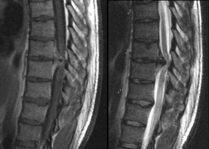
| Herniated T9-10 Disk:
Sagittal MRI scan of
the thoracic spine; (Left) T1-weighted with gadolinium; (Right) T2-weighted. Note
the large disk herniation between the T9 and T10 vertebral bodies, deforming the
spinal cord at that level. Also note the effacement of the CSF both anteriorly
and posteriorly, best seen on the T2-weighted scan (where CSF is bright). In
this case, the spinal cord compression resulted in the leg weakness
and brisk reflexes. Among spinal disorders, herniated disks are very common. Disks are located between the vertebral bodies. They are composed of a thick annulus fibrosis surrounding a jelly like interior known as the nucleus pulposus. The anterior and posterior longitudinal ligaments run along the front and back of the vertebral bones and disks respectively. Disk herniations most often occur posterior laterally, where they compress the exiting nerve root at that level. Straight posterior disk herniations, although less common, may result in direct spinal cord or cauda equina compression. |
Revised
11/23/06
Copyrighted 2006. David C Preston