|
A 57 year-old woman developed radiating pain around her upper torso. Three weeks later, she developed difficulty walking along with bladder incontinence. She also complained of pain between her shoulder blades. |
![]()
![]()
![]()
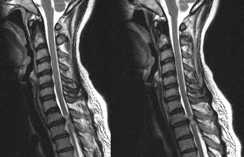
| Herniated T1-2 Disk:
T2-weighted sagittal MRI scans of
the cervical and upper thoracic spine. Note
the large disk herniation between the T1 and T2 vertebral bodies. Also note the effacement of the CSF both anteriorly
and posteriorly. If one looks at the spinal cord, one can see a bright signal
within the cord. This finding occurs either from gliosis or ischemia
from the compressing disk, and portends a poorer prognosis. In
this case, the spinal cord compression resulted in the leg weakness
and bladder incontinence. Among spinal disorders, herniated disks are very common. Disks are located between the vertebral bodies. They are composed of a thick annulus fibrosis surrounding a jelly like interior known as the nucleus pulposus. The anterior and posterior longitudinal ligaments run along the front and back of the vertebral bones and disks respectively. Disk herniations most often occur posterior laterally, where they compress the exiting nerve root at that level. Straight posterior disk herniations, although less common, may result in direct spinal cord or cauda equina compression. |
Revised
11/30/06
Copyrighted 2006. David C Preston