|
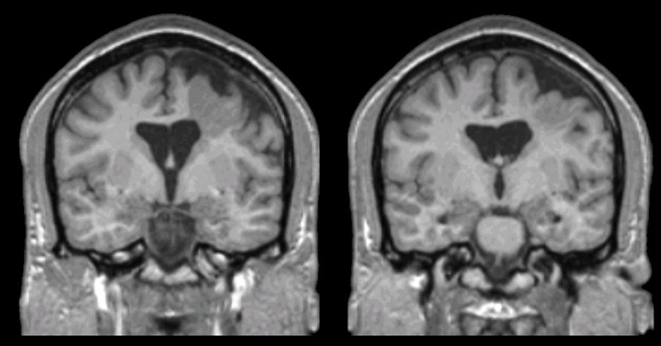
A 22 year-old man with mild mental retardation underwent evaluation for intractable complex partial seizures. |
![]()
![]()
| Heterotopia:
MP-RAGE coronal MRIs. Note the presence of the malformed gray
matter that extends from the left lateral ventricle to the surface of the
left frontal lobe. Also note that the adjacent brain is atrophic,
resulting in an enlarged space between the surface of the brain and
the dura. This is most likely heterotopic gray matter.
However, it is also possible that this is "closed
lip" schizencephaly wherein the two sides of the cleft have fused
together (see the images on schizencephaly,
closed lip and
open lip). Heterotopic gray matter is a developmental malformation wherein some neurons do not migrate to their expected location, instead forming clumps of gray matter, usually in the deep cerebral white matter or lining the walls of the lateral ventricles (periventricular nodular heterotopia). This condition is on the spectrum of neuronal migration disorders. Neurological sequelae may include seizures, mental retardation, learning disabilities and impaired fine coordination. |
Revised
11/30/06
Copyrighted 2006. David C Preston