|
A 55 year-old man presented with a headache, vomiting and a left hemiparesis. |
![]()
![]()
![]()
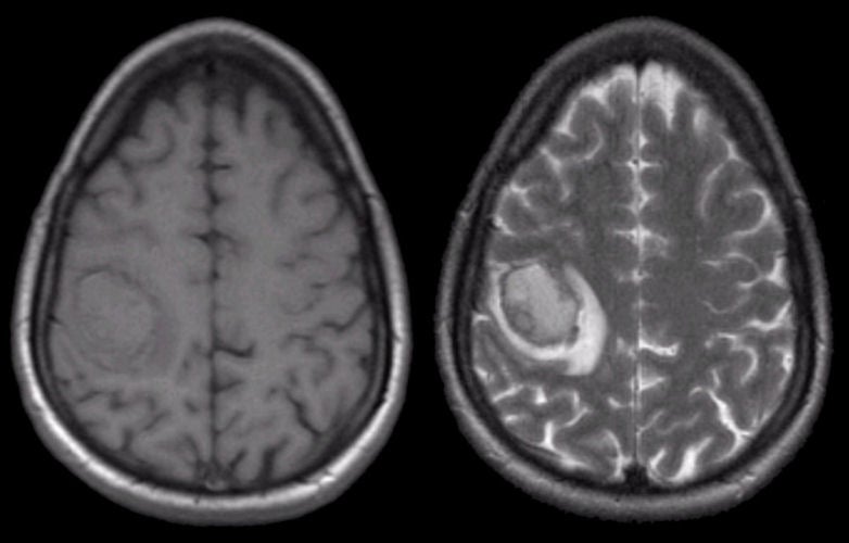
| Hyperacute Intracerebral Hemorrhage: (Left) T1-weighted
axial MRI; (Right) T2-weighted axial MRI. On the T1-weighted scan, note
that there is an abnormality in the right posterior frontal
lobe that is relatively isointense. The same
area on the T2-weighted scan is isointense / hyperintense with a surrounding bright signal.
This is the characteristic picture of a hyperacute (approximately 1 day old) hemorrhage on MRI. In the hyperacute stage, intracellular oxyhemoglobin is isointense / hypointense on T1-weighted and isointense / hyperintense on T2-weighted scans. As oxyhemoglobin changes to deoxyhemoglobin, the signal turns dark both on T1- and T2-weighted scans. This occurs first at the periphery. This process can be seen just beginning to occur on both images. In this case, the hemorrhage was due to hypertension. The findings of blood on MRI are complex and depend on timing. To learn more, review the powerpoint slide show, Blood on MRI: Time-dependent Changes. |
Revised
11/30/06.
Copyrighted 2006. David C Preston.