|
An 11 year-old boy presented with a one month history of headaches. His neurological examination was normal. |
![]()
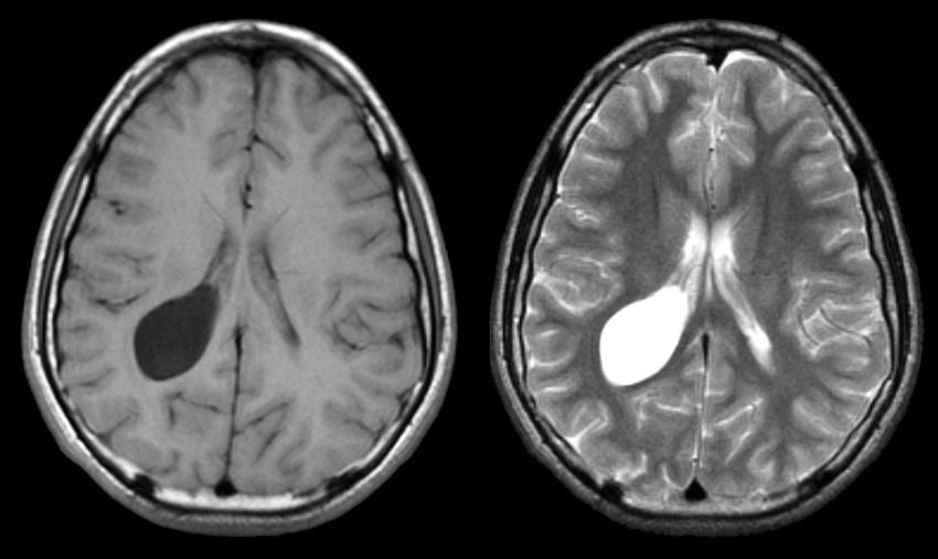
| Intraventricular Arachnoid Cyst:
(Left)
T1-weighted axial MRI; (Right) T2-weighted axial MRI. Note
the large cyst in the atrium of the right lateral ventricle.
The cyst contains fluid, thus it is dark on T1- and bright on
T2-weighted images. Arachnoid cysts are common congenital malformations. They contain fluid but generally do not communicate with the ventricular system. The most common locations are the Sylvian fissure and middle cranial fossa , suprasellar cistern, quadrigeminal cistern, cerebellopontine angle, posterior infratentorial midline, cerebral convexity and interhemispheric fissure. Rarely, these may be found inside a ventricle, as in this case. Most are asymptomatic and found incidentally on brain imaging studies. Rarely, they can enlarge and present as
a mass lesion with focal neurological signs, seizures or hydrocephalus.
If symptomatic, they may be treated with resection/fenestration or
shunting. |
Revised
11/29/06
Copyrighted 2006. David C Preston