![]()
![]()
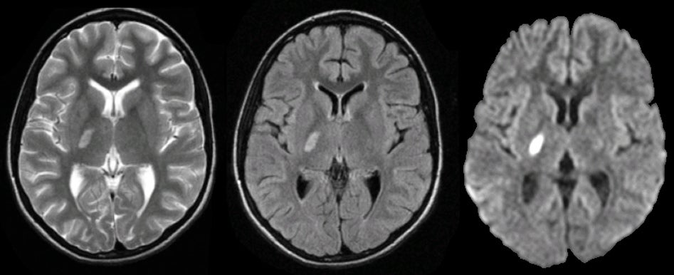
| Lacunar Infarction:
(Left) T2-weighted axial MRI; (Middle) Flair axial MRI; (Right) Diffusion-weighted
axial MRI. Note the
single white matter lesion
in the posterior limb of the right internal capsule. It is seen in
the T2-weighted, Flair, and most prominently in the diffusion-weighted image. This is
a subacute lacunar infarct. Lacunar strokes (also known as small vessel disease) are caused by occlusion of the deep perforating blood vessels. Small vessel disease is most commonly associated with hypertension and diabetes. There are several classic lacunar syndromes, including pure motor hemiparesis, ataxic hemiparesis, clumsy hand-dysarthria (caused by lesions either in the internal capsule or basis pontis) and pure sensory stroke (caused by a lesion in the thalamus). Remember that lacunar strokes are NOT associated with cortical findings, such as aphasia (except rarely), apraxia, neglect, or visual field abnormalities. |
Revised
11/25/06
Copyrighted 2006. David C Preston